AMH Testing for Ovarian Reserve Outside Fertility Contexts
AMH testing reveals declining ovarian reserve years before menopause.
AMH testing measures how many follicles remain in the ovaries at a given point in time, and for years that measurement lived almost entirely inside fertility clinics: IVF planning, PCOS workups, oncofertility consults before chemotherapy. That framing missed something. Declining ovarian reserve is not only a fertility signal, it is a systemic hormonal shift that starts years before a woman's final period, and it shapes symptom timing and long-term health risk whether or not she is trying to conceive. A woman in her early 40s with unexplained fatigue, disrupted sleep, or new anxiety has likely never been offered this test, because nobody told her it applied to her.
What AMH measures and why it behaves differently from FSH and estradiol
Anti-Müllerian hormone comes from granulosa cells, the cells that line small growing follicles from the primary stage through early antral development. Primordial follicles, the dormant ones sitting in reserve, don't secrete it. So AMH is a read on the follicles that are already active and growing, which makes it a reasonable proxy for the size of the pool still available.
What makes AMH clinically useful isn't just what it measures, it's when you can measure it. Secretion runs largely independent of the gonadotropin swings that drive the menstrual cycle. FSH and estradiol rise and fall across each cycle, sometimes sharply, which means timing the blood draw matters enormously for those two. AMH doesn't have that problem. It can be drawn at different points in the cycle and the number means roughly the same thing either way.
FSH tells a messier story. It rises as follicular reserve falls, because the pituitary gland is working harder to push folliculogenesis forward, but FSH also spikes at the LH surge even in women with completely normal reserve. That overlap creates real ambiguity. The Endotext chapter notes that FSH was the first hormone linked directly to ovarian aging, but AMH and antral follicle count now deliver more sensitive, more specific readings. Inhibin B, meanwhile, functions more like a flow gauge, reflecting moment-to-moment follicular activity, while AMH acts like a stock gauge, showing what's left in reserve, a distinction laid out in a 2026 Frontiers in Endocrinology review.
None of this means AMH tells you about egg quality, pregnancy odds, or why a cycle skipped a month. It doesn't, and that limitation matters enough to repeat later in this piece. But for a woman already dealing with irregular cycles, the defining feature of perimenopause, AMH's stability is exactly what makes it interpretable when FSH, drawn at the wrong moment, might not be.
How AMH levels change across the reproductive lifespan
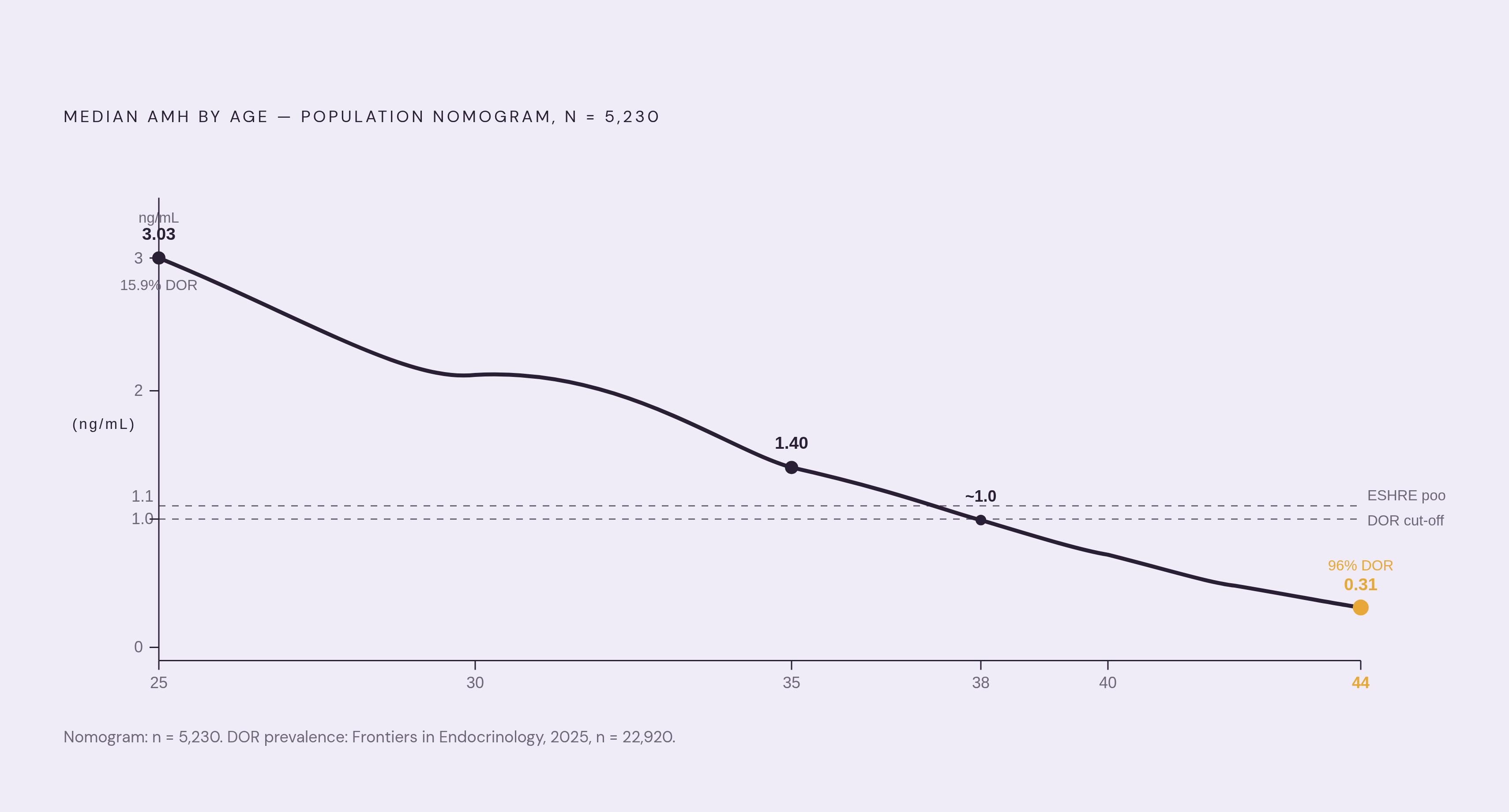
The broad trajectory is well documented. Personalabs reports that AMH is around 3.0 ng/mL in the mid-20s, drifts down to roughly 1 ng/mL by age 40, and is near 0.5 ng/mL by 45. Labs generally treat 1.0 to 4.0 ng/mL as the normal range for reproductive-age women, below 1.0 ng/mL as a possible sign of diminished ovarian reserve, and above 4.0 ng/mL as a marker sometimes associated with elevated follicle count, including in PCOS. The ESHRE Bologna criteria set the poor-response threshold at below 1.1 ng/mL, while ACOG and ASRM define diminished ovarian reserve using below 1.0 ng/mL combined with FSH above 10 mIU/mL.
A more granular picture comes from a population-based nomogram study of 5,230 women. It found the median AMH falling from 3.03 ng/mL at age 25 to just 0.31 ng/mL at age 44. Even the 90th percentile, meaning women on the higher end of reserve for their age, shows a steep floor: 0.78 ng/mL at 25, 0.71 at 30, 0.39 at 35, and just 0.13 by 40. A separate, larger nomogram published in Frontiers in Endocrinology in 2025, drawing on 22,920 women, found median AMH dropping below 1.2 ng/mL by age 36, with the prevalence of diminished ovarian reserve climbing from 15.9% at age 18 to 96% at age 45.
Sit with that last number for a second. Ninety-six percent, by 45. These figures make legible something a lot of women already sense but can't name: "early" perimenopause symptoms in the mid-to-late 30s aren't imagined or premature, they correspond to a real, measurable decline in the follicular pool that's already well underway.
One caveat needs to sit alongside every number in this section. Diminished ovarian reserve has no single, universally accepted clinical definition. Different providers use different combinations of AMH, antral follicle count, FSH, or response to fertility treatment to define it. So no single number from a single test should be read as a verdict.
What a declining AMH signals about perimenopause, not just fertility
A shrinking follicular pool means less estrogen and progesterone production over time, and that's a gradual mechanical fact, not a switch that flips. The hormonal turbulence that defines perimenopause, the erratic cycles, the unpredictable ovulation, is a direct product of a reserve that's getting thinner and less responsive. AMH doesn't just track alongside that turbulence, it captures the underlying driver of it.
A 2026 study on PubMed, covering 10,543 women in a cross-sectional cohort and 2,521 in a longitudinal cohort, found that among women aged 35 to 55, lower AMH percentiles correlated with more severe perimenopausal symptoms (β = 0.12), vasomotor symptoms especially, meaning hot flashes and night sweats. But the level alone wasn't the whole story. The 2026 PubMed study also found that women with lower AMH percentiles experienced more severe symptom progression, which means the rate of change carries information the level alone doesn't.
How much does a low level actually shift the odds? WHOOP's Women's Health Biomarkers data found that an AMH of 0.5 ng/mL carries an odds ratio of 23 for early menopause compared to a level of 2.0 ng/mL. That's a substantial gap for two numbers that might look similarly "low" to someone unfamiliar with the scale.
And the timeline keeps pushing earlier than most people expect. A UVA Health and Flo Health study found that 55.4% of women aged 30 to 35 reported moderate to severe perimenopausal symptoms on the Menopause Rating Scale. That figure is not a typo or a fringe finding; it represents over half of women in that age group. The systemic stakes go well past hot flashes and irregular periods, too: the Endotext chapter notes that estrogen's influence extends to cardiovascular, skeletal, and mental health. So knowing when estrogen production starts declining meaningfully isn't a niche reproductive question, it's closer to a whole-body one.
What this section is not claiming matters just as much as what it is. AMH doesn't predict the exact date of menopause, and it doesn't guarantee a particular symptom path. It signals direction and pace. Destination is a different question.
Where AMH fits within the STRAW+10 staging framework and alongside other hormone markers
STRAW+10, built out of the original Stages of Reproductive Aging Workshop from 2001, gives clinicians a standardized way to stage reproductive aging using menstrual patterns, hormone levels, and antral follicle count together. The framework treats reproductive aging as a gradual, multi-stage process rather than one event, and AMH sits among the named hormone markers alongside FSH, inhibin B, and estradiol.
Mapped onto the stages, the pattern looks like this. In the late reproductive stage, AMH starts declining noticeably even while FSH may still read normal and cycles stay regular, which is precisely the window where AMH tells you something FSH can't yet see. Moving into early perimenopause, what STRAW+10 calls the menopausal transition, cycles start to lengthen or shorten unpredictably, FSH begins rising, AMH keeps dropping, and hormonal output continues to shift. By late perimenopause, cycles become infrequent enough that once 60 days pass without a period, the stage is considered likely, AMH reads very low or undetectable, and estrogen levels become increasingly variable overall.
Timing-wise, perimenopause typically starts 8 to 10 years before menopause itself and runs 4 to 10 years total, with a European average onset age of 47 according to the FSRH's guidance. Early perimenopause tends to last 2 to 4 years, late perimenopause 1 to 3 years, and where a woman's AMH sits corresponds roughly to her position along that stretch.
Why bother tracking AMH at all if FSH is already part of the standard picture? Because FSH is reactive. It reflects the pituitary's response to a feedback loop that's already breaking down, which means by the time FSH is clearly elevated, the reserve behind it has been declining for years already. AMH, by contrast, reflects the reserve directly, in something closer to real time. Inhibin B complements both by showing dynamic, FSH-driven follicular activity, the "flow" to AMH's "stock," though the Frontiers in Endocrinology review notes it's been underused historically because its dynamic nature makes it harder to standardize into a single assay.
None of this is meant to turn a reader into a clinician. The point of laying out STRAW+10 here is to give a woman a map she can locate her own experience on, not to hand her a glossary of terms she'll never need again.
What AMH cannot tell you and where the limits of the test genuinely lie
Start with the biggest misconception first: AMH does not predict fertility. It reflects quantity, not quality. A low result doesn't diagnose infertility, and a normal result doesn't confirm the ability to conceive. Those are separate questions that AMH, by design, doesn't answer.
It also doesn't predict the precise date of menopause for any one woman. It signals the pace of decline rather than a scheduled event on a calendar. And it doesn't diagnose perimenopause on its own, either, since that remains a clinical diagnosis built from symptom pattern and menstrual history, with hormone levels serving as supporting evidence rather than the whole case.
Then there's the issue of different labs using different AMH assays, which is less discussed but genuinely significant. Different labs use different AMH assays, and those assays can produce different absolute values for the same blood sample. Reference ranges vary from lab to lab and don't always match the population nomograms used in research studies. Standardization across assays and age-adjusted reference ranges remains an unresolved issue in the field.
Hormonal birth control complicates things further. It can temporarily suppress AMH, making reserve look lower than it actually is, which matters a great deal for any woman currently on the pill, a hormonal intrauterine device, or another hormonal method who gets tested and sees a number lower than expected.
The Endotext chapter from December 2025 states that no single AMH result is definitive on its own. Findings need context, and often need to be repeated or paired with other markers before they mean much. Combine that with the fact that diminished ovarian reserve has no single agreed-upon clinical definition, and the conclusion is unavoidable: AMH works best as a directional signal, tracked over time, read next to other markers and a woman's actual clinical picture. Not as a verdict delivered by one number on one day.
How to interpret an AMH result outside a fertility clinic, reading it as a hormonal health signal
The same number means different things to different women, and that context has to come first. A 0.9 ng/mL result lands very differently for a 38-year-old noticing her cycles shortening than it does for a 32-year-old mapping out an IVF cycle. Same test, same lab, entirely different meaning.
A result between 1.0 and 4.0 ng/mL suggests reserve sitting within population norms for reproductive-age women, though that doesn't rule out perimenopausal symptoms if estrogen is already fluctuating underneath a still-normal AMH. A result under 1.0 ng/mL, paired with irregular cycles or vasomotor symptoms like hot flashes, lines up with accelerated ovarian aging. That's a signal to track closely, not a number to panic over.
The direction between two numbers affects how the decline should be interpreted, more than either number alone can. The 2026 PubMed study cited earlier found that women with lower AMH percentiles experienced more severe perimenopausal symptoms, a pattern that's only visible with more than one data point sitting side by side. A single test is a photograph. Two tests, months or years apart, start to look like a trend line.
Reading AMH alongside other markers sharpens the picture further. A concurrent decline in AMH with a rise in FSH offers a strong, convergent signal of advancing reproductive aging. AMH low alongside fluctuating estradiol fits the profile of late reproductive stage or early perimenopause. AMH low while cycles are still regular suggests an early transition to monitor rather than a conclusion to draw yet.
Maybe the most useful way to think about a low AMH result is as an opening rather than a closing. For a woman dealing with unexplained fatigue, mood shifts, or disrupted sleep, a low AMH gives a clinician a concrete starting point to investigate hormonal causes, instead of defaulting to stress or anxiety as the explanation. But a number without someone qualified to place it in a fuller clinical picture isn't a health strategy on its own, which is why clinician review matters here as much as the test itself. AMH can now be ordered without a doctor's visit through direct-access lab services, with no fasting or cycle-day timing required. Getting the number is the easy part. Knowing what to do with it actually requires expertise.
Why tracking AMH over time reveals what a single test cannot
A reading of 0.8 ng/mL at age 42 means something entirely different depending on what came before it. If a prior test at 40 showed 1.8 ng/mL, that's a steep, fast decline. If it showed 0.9 ng/mL, that's a slow, expected drift. One data point can't tell the difference. Only two can.
That distinction isn't hypothetical, it's the specific finding the 2026 study (10,543 women cross-sectional, 2,521 longitudinal) was built to catch. Women with lower AMH percentiles showed more severe perimenopausal symptoms, a pattern invisible in cross-sectional data alone and only detectable by following the same women across multiple tests over time.
So what should a reader actually take from that? A single AMH result, taken in isolation, functions more like a weather report than a climate forecast. It tells you the conditions today. It says very little about the season ahead unless there's a second reading, and ideally a third, to compare it against. That's not a flaw in the test, it's simply how a stock measurement works. The number itself is stable and easy to read on any given day, but its real value appears only in the shape of the curve it draws over months and years, not in the isolated dot it leaves behind on any one visit.
Sources
- Ovarian Reserve Testing
- frontiersin.org
- AMH Blood Test — Check Your Ovarian Reserve Online
- Age-stratified anti-Müllerian hormone (AMH) nomogram: a comprehensive cohort study including 22.920 women - PMC
- ncbi.nlm.nih.gov
- whoop.com
- Developing anti-Müllerian hormone as an ovarian reserve biomarker
- Challenges in Measuring AMH in the Clinical Setting