DEXA Scan vs Blood Biomarkers for Bone Health in Perimenopause
Blood markers track bone loss years before DEXA scans catch the decline.
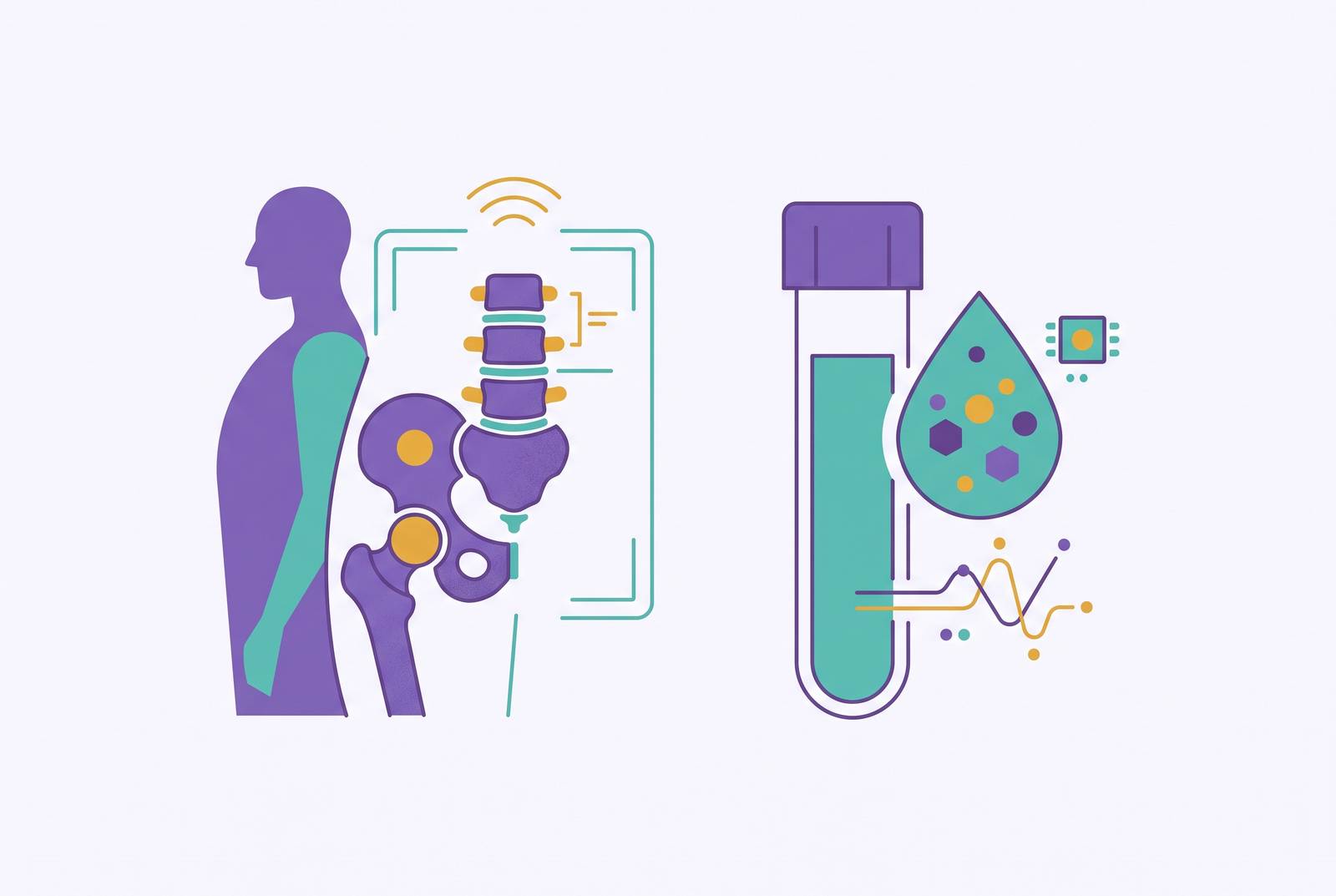
Bone loss during perimenopause starts years before most women get anywhere near a screening, and it moves in near total silence. Two tools track it: DEXA scans and blood biomarkers. They measure different things on different timelines, and relying on just one leaves real gaps in the picture. Perimenopause starts around age 47 on average and runs anywhere from two to ten years, a long stretch for a process that gives almost no warning until something breaks. Knowing which tool catches which kind of change, and when, is what separates seeing this coming from finding out after a fracture.
The standard approach, wait for symptoms, then screen, gets the timing backwards. Perimenopause is when resorption starts outrunning formation, and this imbalance causes a meaningful amount of bone to be gone before a diagnostic report ever flags osteoporosis. By the time the scan flags a problem, the problem has had years to compound.
What is happening inside bone during the hormonal transition
Bone is living tissue, broken down and rebuilt continuously by two cell types working against each other: osteoclasts dismantle it, osteoblasts rebuild it. Density at any given moment is just the scoreboard for that fight.
Estrogen's job is to keep osteoclast activity in check. As estrogen falls through perimenopause, that brake loosens, resorption speeds up, and formation can't keep pace. Bone mineral density starts to slide. For years that was treated as the whole story: falling estrogen, rising resorption, end of explanation.
But a second actor gets far less attention than it should. FSH, follicle-stimulating hormone, rises sharply in late perimenopause and appears to drive osteoclast activity on its own, apart from whatever estrogen is doing. Research published in an MDPI journal on the menopause transition found that each doubling of FSH during the transition tracks with a modest but real additional annual decline in bone mineral density at the femoral neck and lumbar spine, and that compounds over several years. Its effect on bone often appears in remodeling changes before estrogen has dropped low enough to cause hot flashes or night sweats, so a woman can feel, by every outward measure, hormonally normal while her bone remodeling has already shifted underneath her. If FSH were simply a stand-in for low estrogen, restoring estrogen should push FSH back to baseline. It often doesn't: estrogen therapy frequently fails to normalize FSH after menopause, and bone loss can continue anyway, which points to FSH working through its own pathway rather than just narrating estrogen's absence.
Progesterone is a third thread, tied to ovulation rather than hormone level alone. The PeKnO Study found that a marked decline in ovulatory rate during perimenopause tracks with faster bone density loss, even in women whose estrogen still looks fine on paper. Anovulatory cycles mean little to no luteal progesterone, and that gap seems to matter for bone well before estrogen has meaningfully dropped.
Estrogen, FSH, and progesterone via ovulation together partially explain why roughly a quarter of women fall into what researchers call the "fast bone loser" category during this transition, losing density fast enough to carry real elevated risk for osteoporosis later. No single hormone number and no single scan can account for three mechanisms running on three different schedules. That is the actual reason this can't be settled with one test.
What a DEXA scan measures and what its numbers mean
DEXA, dual-energy X-ray absorptiometry, is a low-radiation scan that measures bone mineral density, usually at the hip and spine, the two sites most predictive of fracture risk. Hip fracture carries the heaviest long-term cost to mobility and independence. That is why those two sites get the attention.
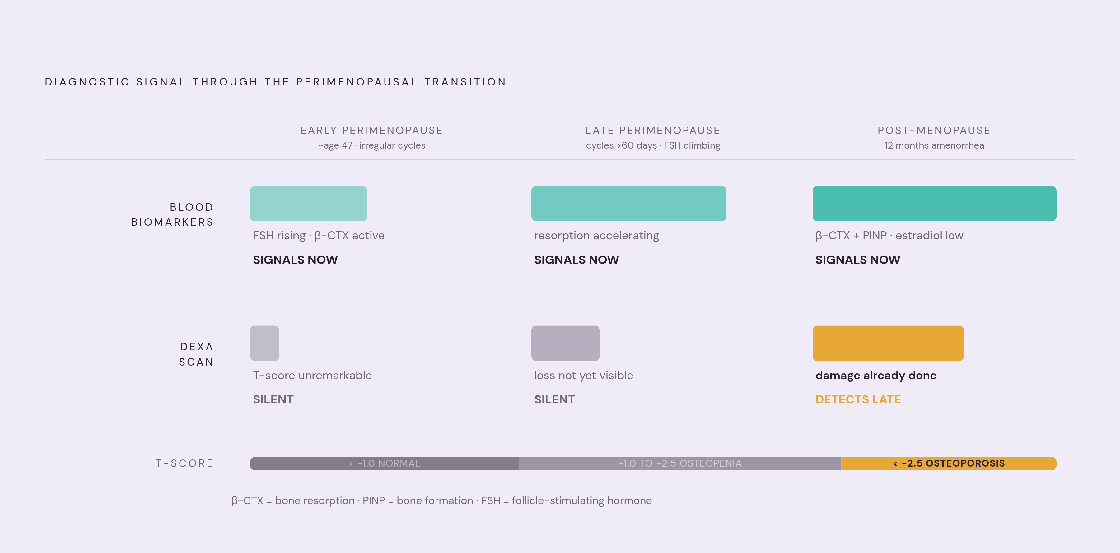
The result comes back as a T-score, comparing a woman's bone density against the average peak bone mass of a healthy 30-year-old of the same sex. Normal sits above -1.0, osteopenia runs from -1.0 to -2.5, osteoporosis is below -2.5. That number says where she stands today against a reference group. It says nothing about how she got there, and nothing about where she's headed.
A Z-score does different work: it compares bone density against peers of the same age and sex rather than a fixed 30-year-old benchmark, and that age-adjusted view catches unexplained loss in younger women that a T-score would miss.
What DEXA does, it does well. The measurement is objective, standardized, and reproducible on the same machine over time, and it remains the clinical gold standard for diagnosing osteopenia and osteoporosis. Treating a single T-score as the whole conversation stops the number from doing its job. Read alongside age, genetic background, hormonal status, body composition, and lifestyle, that same score means far more. It is a starting point.
Why DEXA screening timing under current guidelines may not serve perimenopausal women
Major clinical guidelines put DEXA screening at age 65 for average-risk women, with rescreening covered roughly every two years under Medicare. Medicare covers a scan every other year past that age. Women with early or surgical menopause, a family history of hip fracture, long-term corticosteroid use, or a prior fragility fracture often get screened earlier. Even that expanded list leaves out a large group of perimenopausal women losing bone quietly without meeting any of those criteria.
Age 65 is the wrong number for a process that does not wait for it. Net bone loss begins gradually in early adulthood, picking up speed through the 40s and 50s, exactly when perimenopause is unfolding. Standard screening arrives a decade or more after the process it is meant to catch has already started. Women can lose a substantial share of bone density in the years following menopause, and that decline is already in motion well before the final period ever arrives. Waiting for the guideline age means watching the scan confirm a loss that finished happening years earlier.
Insurance coverage is not as locked to age 65 as the default guideline suggests, either. Earlier scans may qualify under certain documented clinical conditions, which can open a real path for perimenopausal women who know to ask their clinician. Functional and longevity-focused medicine pushes further still: Parsley Health's clinical position calls for at least one full-body DEXA after age 30, ideally with full body composition every one to two years starting around 35, framed not as a diagnostic trigger but as a baseline laid down before the losses pick up speed.
That baseline is what makes a later scan mean anything. A DEXA at 48 with nothing to compare it to is a snapshot. A DEXA at 48 measured against one from age 38 is a trajectory, and trajectory, not a single point, is what actually predicts what comes next. Even with earlier scanning in place, a gap remains: a DEXA taken today only shows the accumulated result of everything that has already happened. It cannot say how fast the loss is happening right now, or which hormonal force is driving it. That is a job for a different kind of data.
What blood biomarkers reveal that a DEXA cannot show
Two categories of blood markers matter, and they answer different questions. Hormone markers, FSH, LH, estradiol, progesterone, show where a woman sits in the transition and which hormonal forces are actively working on bone at that moment. Bone turnover markers (BTMs) are biochemical signals of resorption and formation happening in real time. A 2026 evidence-based narrative review indexed in a major medical literature database (42085078) found that pairing bone mineral density with BTMs such as β-CTX (a resorption marker) and PINP (a formation marker) sharpens fracture risk assessment beyond what density alone can offer.
BTMs catch activity a DEXA physically cannot register. A woman can carry a T-score sitting comfortably in the normal range while her resorption markers run hot, and that combination functions as an early warning that a future DEXA is likely to look worse than this one. Density alone can't supply that warning, since it only reflects what has already accumulated, never the rate at which change is happening right now.
Hormone markers explain the why behind that rate. FSH elevation in late perimenopause tracks with faster bone remodeling independent of estradiol, so watching FSH over time helps pin down when that particular driver is most active. A cross-sectional study in Scientific Reports reported average FSH of 44.84 IU/L in perimenopausal women, climbing to 74.07 IU/L postmenopause, a gradient that lines up with rising bone risk across the transition. Those are population averages, not personal reference ranges, but the direction is unmistakable. Estradiol moved the opposite way in the same study, averaging 65.76 pg/mL perimenopausal versus 38.08 pg/mL postmenopause, tracking the loss of estrogen's protective effect as the transition wears on.
Progesterone belongs here too, through the same mechanism the PeKnO Study flagged: anovulatory cycles, meaning low or absent luteal progesterone, track with faster bone loss even while estrogen numbers still look fine on paper. Vitamin D, calcium, and inflammation markers round out a real bone-health workup, since deficiencies in any of them drive loss on their own separate track, apart from the hormonal cascade.
Blood biomarkers describe what's happening now and why. DEXA describes what has already piled up. Relying on one instead of both means losing half the story, and that lost half would have caught the problem in time to act.
The interpretation challenge: why neither tool works well as a one-time reading
A single T-score has an obvious blind spot. It says where a woman stands against a reference population at that exact moment. It says nothing about whether she is losing bone slowly or rapidly, or whether her trajectory is improving or worsening. Two women can carry identical T-scores while on completely different paths, one stable, one in free fall, and the scan alone cannot tell them apart.
Hormone markers have a worse version of the same problem. FSH and estradiol swing substantially within a single cycle and across cycles during perimenopause, so a single blood draw can falsely reassure or falsely alarm depending purely on where in the cycle it lands. Age and symptom pattern deserve more clinical weight than any one lab value. A 47-year-old with irregular cycles, hot flashes, and broken sleep should not be told she is fine because one FSH draw came back "not high." A single number rarely tells the truth about a transition this noisy, and treating it as decisive is the mistake most likely to delay real intervention.
Bone turnover markers need the same caveat. A single elevated β-CTX reading means far more measured against an earlier baseline than it does sitting alone with nothing behind it.
Longitudinal tracking is what actually earns its keep here. A rising FSH trend tracked over time, paired with falling estradiol and a pattern of anovulatory cycles showing up in progesterone, tells a coherent story no single blood draw can tell. A follow-up DEXA after baseline turns a static number into a rate, and that rate, not the number itself, is what predicts where bone density goes next. BTMs measured at intervals can show whether an intervention, a lifestyle change, a supplement, hormonal support, is actually slowing resorption well before the next DEXA would confirm it.
None of this replaces clinical judgment. A T-score, an FSH value, a β-CTX level: none of these numbers turns into a decision by itself. Each needs a clinician's read to become useful, and each does its best work tracked in sequence over time, not filed away as a one-off check and forgotten.
Using both tools together across the perimenopausal timeline
Early perimenopause looks different from late perimenopause, hormonally and structurally, and the tools should shift with it rather than getting deployed the same way at every stage.
In early perimenopause, when cycles are just starting to go irregular and FSH and estradiol are still bouncing around, blood biomarkers carry the most weight. FSH, LH, estradiol, and progesterone establish roughly where a woman sits in the transition and flag anovulatory patterns, while vitamin D and relevant inflammation markers catch modifiable drivers of bone loss unrelated to reproductive hormones. If a baseline DEXA exists from around age 30 to 35, this is the point to run a comparison against it. If no baseline exists yet, this is a reasonable window to create one, not to diagnose osteoporosis, but to lay down the reference point that makes every future scan mean something. BTMs fit in here too, since they can flag accelerating resorption while the DEXA number itself still looks unremarkable.
Late perimenopause, once cycles stretch past 60 days apart and FSH climbs more consistently, is where the hormone-driven, estrogen-independent bone loss described earlier tends to be most active. A DEXA comparison against baseline carries more weight at this stage. For women never scanned before, the case for screening earlier than 65 gets stronger here, especially with documented low estradiol on record to support coverage. If resorption markers run elevated at this point, shortening the interval to the next DEXA makes more sense than waiting the full standard interval.
What the two tools accomplish together is something neither manages alone. DEXA supplies the structural outcome: current bone mineral density and how it has shifted since the last scan. Blood biomarkers show which hormonal forces are active and reveal how fast remodeling is running, information measured through hormone levels rather than through that structural number. Combined, a clinician can tell apart a woman whose T-score is stable because her bone turnover genuinely runs slow from a woman whose T-score looks fine today but whose biomarkers are already forecasting meaningful loss by the time the next scan comes around.
Women do not need to wait until 65, or a fracture, or a formal osteoporosis diagnosis to start building this picture. Knowing the qualifying criteria that unlock earlier DEXA coverage, and asking for a full hormone panel instead of a single FSH check, are steps available now, not later. DEXA and blood biomarkers answer different questions about the same skeleton, and treating them as interchangeable, or leaning on either one alone, leaves the picture incomplete during the exact window when catching the pattern early still has room to change what happens next.