Imaging Tests That Are and Are Not Useful in Perimenopause Workups
Blood tests and symptom patterns diagnose perimenopause; imaging has one documented role.
⟦H1⟧
Perimenopause is diagnosed with hormone patterns and symptom tracking, not with a scan. Imaging has exactly one well-supported role in this workup, bone density measurement, and gets ordered far beyond that role more often than it should. What follows lays out where the evidence actually supports a scan, and where it just adds noise, cost, and delay to a diagnosis that blood work and clinical pattern recognition were already built to make. Imaging Tests That Are and Are Not Useful in Perimenopause Workups.
Diagnosing perimenopause on symptoms and hormones, not anatomy
Perimenopause is a multi-year transition marked by fluctuating hormone levels. It is not an event a scan can catch in a single frame. The clinical framework built to track it, STRAW+10, stages the process using menstrual pattern alongside hormone markers like FSH, AMH, estradiol, and inhibin B. None of that is something a picture can capture: an ultrasound cannot measure a hormone level, and it certainly cannot measure a pattern that unfolds across months or years.
The ESE Clinical Practice Guideline, backed by the Endocrine Society, the European Menopause and Andropause Society, and the British Menopause Society, treats menopause as a continuum, with perimenopause and postmenopause as connected stages rather than isolated events.
NICE guidance draws a sharper line. For adults 45 or older presenting with vasomotor symptoms and cycle changes, the diagnosis is clinical. No lab work is required to confirm it, and by extension, no imaging is required either. That single recommendation carries real weight. A scan ordered at that stage is not filling a diagnostic gap, because none exists to fill, and clinicians who order one anyway are treating a functional, hormonal transition as though it were a structural question.
What blood-based testing covers in a perimenopause workup
FSH is the test almost every woman asks for by name, but it is least suited to standing alone. Under STRAW+10, a single blood draw can be in the menopausal range one week and drop back into a reproductive-years range on a different draw taken not long after, which makes one result close to meaningless. There is no universal cutoff either: different numbers cited across the literature describe different stages of the transition, not one threshold that applies to every woman at every point.
Clinical staging leans on menstrual pattern together with hormone markers like FSH, AMH, estradiol, and inhibin B. At 45 and older with typical symptoms, testing FSH adds little given the clinical picture already on the table. Between 40 and 45, it might function as supporting evidence. Under 40, the workup tightens considerably: both NICE NG23 and the ESE guideline call for two samples drawn four to six weeks apart. One detail trips up a lot of workups here: combined hormonal contraception invalidates FSH as a menopausal marker entirely, so testing it while a patient is on the pill produces a number that means nothing.
Estradiol carries a different kind of weight. A 2025 cross-sectional study in Frontiers in Endocrinology identified a specific threshold, 62.7 pmol/L, as predictive of perimenopausal bone loss https://bluepointmedgroup.com/the-perimenopause-workup-what-a-primary-care-visit-actually-checks/. Medicare generally covers DXA every other year for women over 65.
AMH has quietly become one of the more interesting markers in the whole picture. It falls as ovarian reserve declines, often before FSH starts climbing, which makes it useful for tracking the trajectory of the transition, though it cannot diagnose perimenopause on its own or pin down when menopause will land.
TSH belongs in this workup for a different reason. It is not a perimenopause marker, it functions as a rule-out test. Fatigue, mood shifts, irregular cycles, and temperature dysregulation occur in both thyroid disease and perimenopause, and TSH testing rules out the former. The USPSTF does not recommend routine thyroid screening for asymptomatic adults, but once someone is symptomatic, which describes the overwhelming majority of women walking into this workup, checking TSH and free T4 is the right move. Cardiovascular risk tracking matters here too: significant estradiol decline across the transition may contribute to adverse changes in LDL-C and apolipoprotein B. SWAN longitudinal data showed that every 50% decrement in AMH in premenopause and early perimenopause was associated with 0.14%.11%, linking AMH to bone loss risk.
No imaging test diagnoses perimenopause (and why that matters clinically)
NICE states this outright: for people 45 or older with typical symptoms, no single hormone test or imaging scan should be used as a standalone diagnostic tool. That is about as unambiguous as clinical guidance gets, and the fact that the guideline committee felt the need to spell it out says something about how often the rule gets ignored in practice.
Perimenopause is a hormonal and symptomatic transition, and no anatomical finding, no organ shape or tissue texture, confirms that someone has entered it. Ovaries do not develop a signature perimenopausal appearance on ultrasound, and the uterus does not undergo a structural change marking the passage into this stage. Imaging visualizes structure: organ size, tissue density, the presence of a mass or a pocket of fluid. Perimenopause is a functional endocrine state instead, defined by fluctuating hormone levels, a shrinking follicular reserve, and ovulatory patterns that shift unpredictably from one cycle to the next. Those are two different categories of information, and no amount of image resolution closes that gap.
So what actually happens when a clinician orders a pelvic ultrasound or an MRI specifically to diagnose perimenopause? The scan is not following an evidence-based protocol, and it is not answering the question it was ordered to answer, even if it happens to turn something up along the way. That gives women a genuinely useful follow-up question for the exam room: what specifically is this looking for? If the honest answer amounts to "ruling out perimenopause," the workup has drifted off course.
DXA and bone density: the one imaging test with a documented role in the perimenopause transition
Bone loss is a direct downstream consequence of the same hormonal shift driving everything else, not a side issue that happens to coincide with perimenopause. It is a direct downstream consequence of the same hormonal shift driving everything else. Estrogen keeps bone formation and bone resorption in balance, and as estrogen declines, that balance tips hard toward resorption while formation slows. Data from the Study of Women Across Nations found that women can lose 5% to 10% of bone mineral density in both cortical and trabecular bone during late perimenopause and the first years after menopause, the fastest rate of bone loss across a woman's entire lifetime https://www.ccjm.org/content/87/4/205.
DXA fits into this picture, but its job needs to be stated precisely. It does not diagnose perimenopause. It measures bone mineral density and estimates fracture risk as estrogen falls. That is the one genuinely evidence-backed reason to order imaging during this window, and it sits in a different category entirely from every scenario described above.
Current U.S. guidance from the 2025 USPSTF Recommendation Statement calls for screening women 65 and older, a B recommendation, extended to postmenopausal women under 65 who carry certain risk factors. Medicare generally covers DXA every other year once a woman passes 65. Other organizations, including the Bone Health and Osteoporosis Foundation, recommend starting scans as early as age 50 for women and men who carry fracture risk factors.
The gap between 50 and 65 is where the guidance actually falls short. Women in their late 40s and early 50s, right in the thick of perimenopause, may already be accumulating meaningful bone loss well before routine screening guidelines say they qualify for a scan. Other organizations and the Bone Health and Osteoporosis Foundation recommend scans beginning as early as age 50 for women and men with fracture risk factors, rather than waiting on an age cutoff that was never built with perimenopause bone loss in mind.
When pelvic ultrasound and other imaging are legitimately ordered alongside a perimenopause workup
Imaging earns its place in this workup only when it answers a specific structural question, never when it is asked to confirm a hormonal one. That distinction organizes everything in this section.
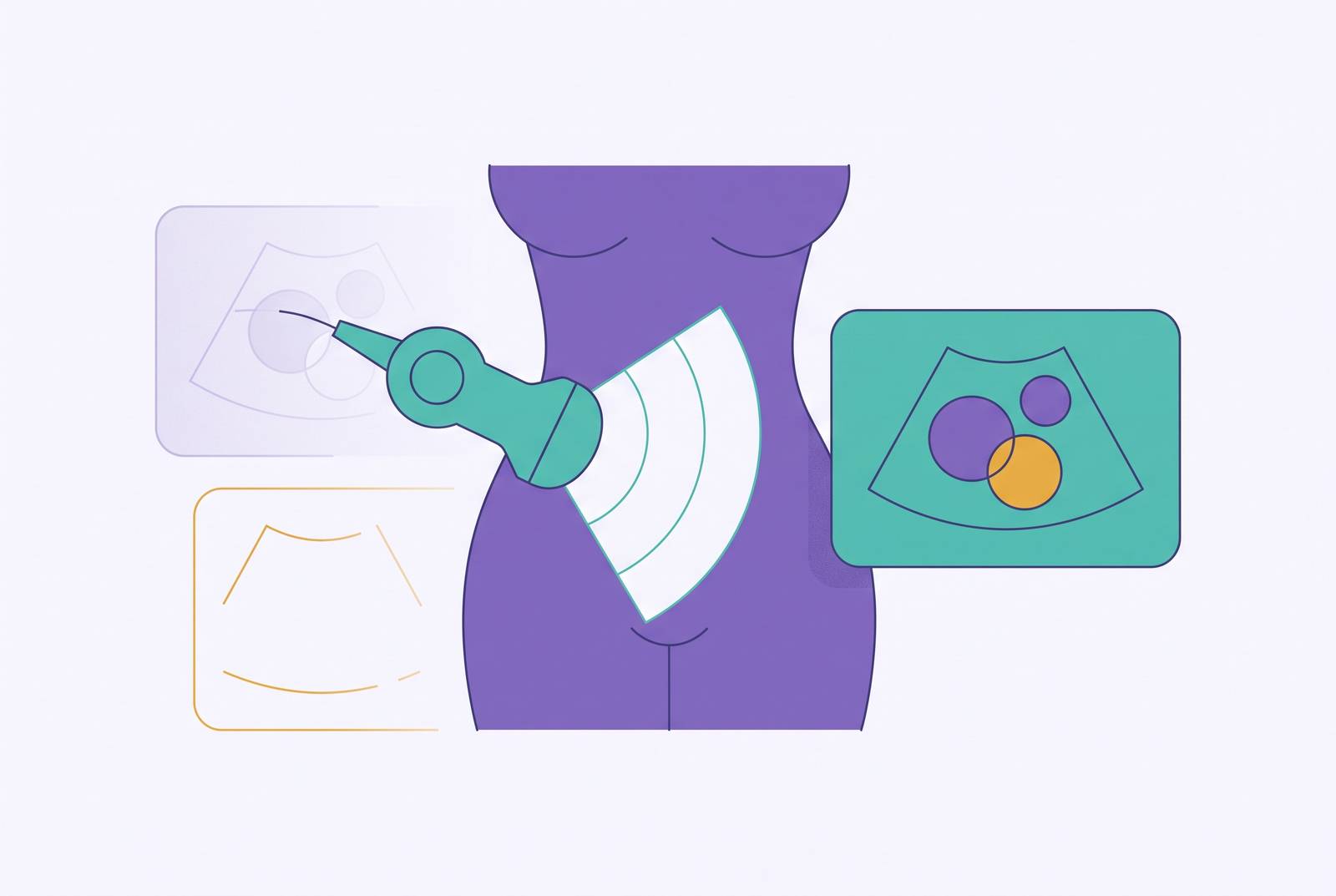
Abnormal uterine bleeding is the clearest case. Heavy and irregular bleeding occurs more often during the menopausal transition, and ACOG defines heavy bleeding with concrete markers: lasting more than seven days, soaking through a pad or tampon every hour for several hours running, or passing clots the size of a quarter or larger. When bleeding meets that threshold, a clinician-guided ultrasound or endometrial evaluation is the right call, aimed at ruling out fibroids, polyps, or endometrial hyperplasia. That scan asks what is causing the bleeding. It does not ask whether the patient is perimenopausal, and confusing the two questions is exactly the mistake to avoid.
Transvaginal ultrasound also shows up in fertility contexts through antral follicle count, or AFC, which appears in STRAW+10 alongside AMH and inhibin B as an ovarian reserve marker. AFC gets used heavily in IVF settings to estimate likely egg yield, but it predicts natural fertility or the timing of menopause poorly. It has no real place in a routine primary care perimenopause workup. It becomes relevant specifically when reproductive planning enters the conversation, a narrower use case than general symptom evaluation.
Premature ovarian insufficiency, or POI, carries its own expanded workup. The ESE guideline notes that patients with POI may go through karyotyping and further genetic testing. POI affects an estimated 1% to 3% of women and comes with elevated risk for osteoporosis, cardiovascular disease, cognitive decline, and depression, which raises the stakes for a thorough evaluation considerably https://www.contemporaryobgyn.net/view/new-guidelines-on-menopause-and-perimenopause-management-released. Fracture risk and cardiovascular risk assessments need to happen before starting hormone replacement therapy in POI patients specifically, given how much higher the stakes run for this younger group.
Why imaging is still over-ordered in perimenopause workups
Perimenopause remains chronically under-diagnosed, and under-explained to the women living through it. A 2025 study in npj Women's Health that looked at 4,432 U.S. women found symptoms appearing far earlier than most people, patients and clinicians alike, tend to expect https://bluepointmedgroup.com/the-perimenopause-workup-what-a-primary-care-visit-actually-checks/. More than a quarter of women between 30 and 35 who saw a doctor for symptoms walked out with a perimenopause diagnosis https://bluepointmedgroup.com/the-perimenopause-workup-what-a-primary-care-visit-actually-checks/. That is a much younger population than the standard mental model accounts for, and it raises an obvious question: if the diagnostic window opens earlier than assumed, how much of the imaging ordered on women in their 30s is aimed at the wrong problem entirely https://www.anylabtestnow.com/blog/menopause-and-perimenopause-which-lab-markers-help-inform-your-plan/?
When a diagnosis feels murky, imaging often gets ordered defensively, as a way to rule out "something structural," even when the answer sitting in front of the clinician is hormonal and calls for blood testing and clinical judgment instead of a scan. That reflex is the wrong move, and it carries real costs. It delays the diagnostic pathway that would actually produce an answer: hormonal evaluation and longitudinal symptom tracking. It adds financial burden to a workup that gains no diagnostic clarity from the extra test. It produces incidental findings that spin off into further workups nobody needed. And it reinforces the exact confusion this piece opened with, women left unsure what perimenopause even is or how it gets properly diagnosed.
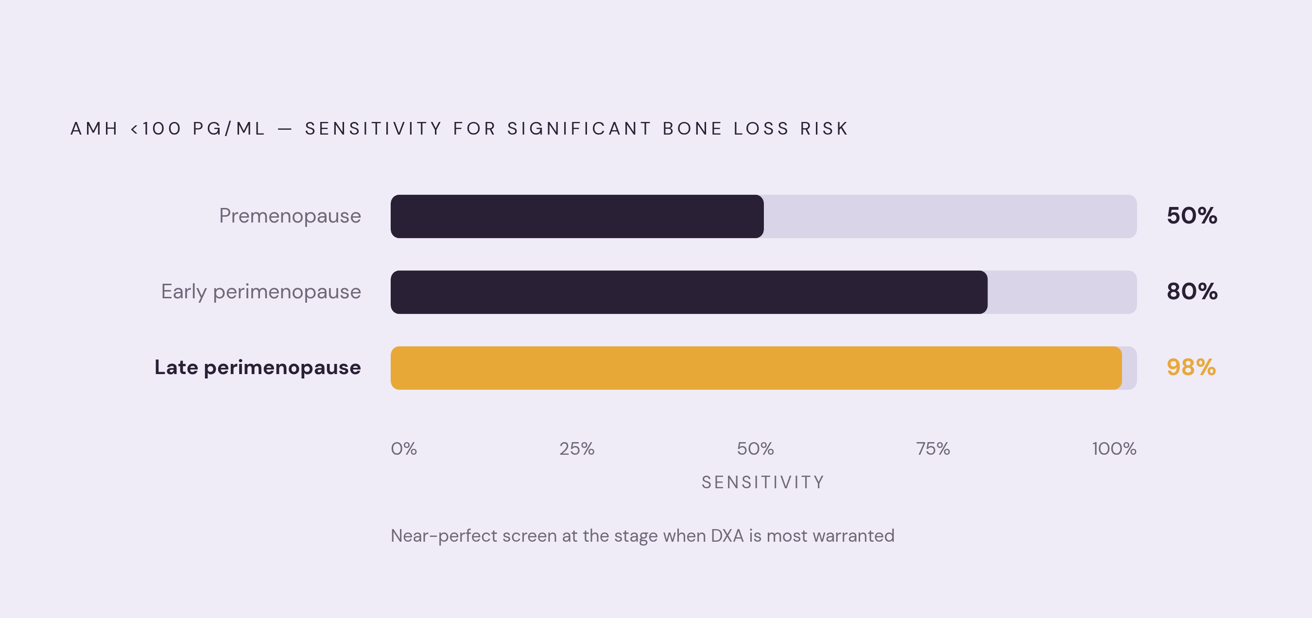
The POI story makes the stakes concrete. The ESE guideline notes that delayed diagnosis of POI carries increased risk of osteoporosis, depression, cognitive decline, cardiovascular disease, and sexual dysfunction. Every one of those risks traces back to a missed endocrine picture, not to a failure of imaging: a scan was never built to catch what blood work and clinical pattern recognition are built to catch. Imaging has a real, evidence-backed job in a handful of specific scenarios, bleeding that needs a structural explanation, bone density that needs a number attached to it, POI that needs genetic follow-up. Outside those lanes, the diagnostic work belongs to hormones and symptoms, and asking a scan to do that job just slows everyone down. Every 50% decrement in AMH level in premenopause and early perimenopause was associated with a 0.14% per year faster decline in lumbar spine BMD over the following 3 to 4 years https://pubmed.ncbi.nlm.nih.gov/35373854/. Every 50% decrement in AMH level in premenopause and early perimenopause was associated with a 0.11% per year faster decline in femoral neck BMD over the following 3 to 4 years https://pubmed.ncbi.nlm.nih.gov/35373854/. AMH below 100 pg/mL will detect women at risk of significant bone loss with sensitivity of 50% in premenopause https://pubmed.ncbi.nlm.nih.gov/35373854/. AMH below 100 pg/mL will detect women at risk of significant bone loss with sensitivity of 80% in early perimenopause https://pubmed.ncbi.nlm.nih.gov/35373854/. AMH below 100 pg/mL will detect women at risk of significant bone loss with sensitivity of 98% in late perimenopause https://pubmed.ncbi.nlm.nih.gov/35373854/. The study included 25 perimenopausal women and 25 premenopausal women https://www.medrxiv.org/content/10.1101/2025.03.20.25324344.full.pdf.