Thyroid Panel Scope in Women's Preventive Testing
Standard thyroid panels miss autoimmune disease and free hormone levels in midlife women.
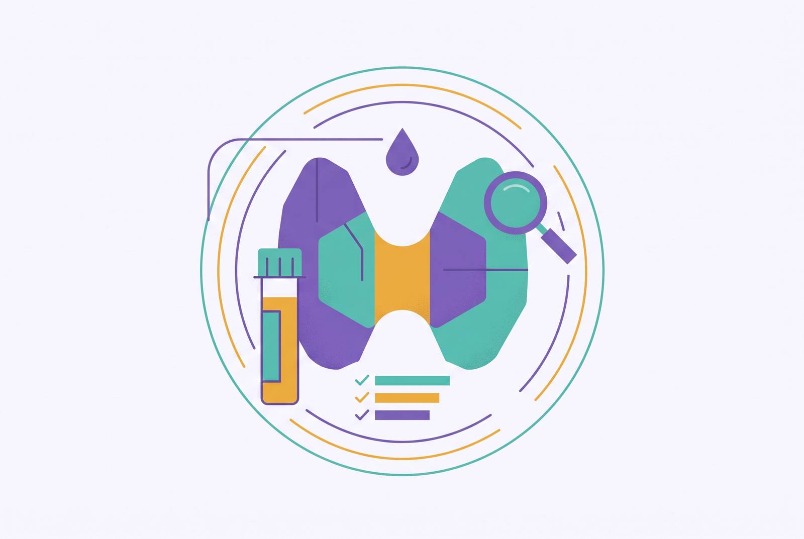
Thyroid dysfunction and perimenopause produce nearly identical symptoms, and most panels are not built to tell them apart. Fatigue, brain fog, mood shifts, weight change, hair thinning, temperature sensitivity: all of it appears in both conditions, so a thyroid problem gets waved off as "just perimenopause" for years, and a hormonal transition gets treated as a thyroid case that never quite resolves. The failure is testing the wrong things, or the right things at the wrong resolution, not a lack of testing. It is testing the wrong things, or the right things at the wrong resolution, and this piece works through why that overlap exists biologically, what a minimum panel misses, what a fuller one adds, and how to build a thyroid-inclusive panel that answers real questions instead of manufacturing new ones.
How common thyroid dysfunction is in midlife women
A woman's lifetime risk of developing a thyroid condition is estimated at around 1 in 8, common enough that any clinician working with midlife women should expect to see it regularly. It is common enough that any clinician working with midlife women should expect to see it regularly, not stumble on it as a surprise.
Prevalence does not sit flat across the reproductive lifespan, either. It climbs specifically during perimenopause and the years right after menopause. The European Menopause and Andropause Society's position statement on thyroid disease and menopause notes that subclinical hypothyroidism is common during the menopausal transition itself. A large cross-sectional study of Korean women aged 40 and older, published in Thyroid in 2022, backs this up at the population level: both overt and subclinical hypothyroidism showed up more often in the late transition and postmenopausal stages, with the authors highlighting the late menopausal transition as the window to watch.
None of this means every abnormal thyroid result demands treatment, and this is where a lot of testing conversations go wrong. Research has shown that a meaningful share of people with subclinical hypothyroidism revert to normal thyroid function on their own, no intervention required. That finding should not talk anyone out of testing. It should inform what happens after the result comes back: a single borderline TSH is information to weigh.
So what does this add up to? Thyroid dysfunction is common enough to expect in a meaningful minority of this population, not some incidental finding that occasionally turns up in a perimenopausal woman's labs. It is common enough to expect in a meaningful minority of this population, and it peaks in prevalence at exactly the moment when symptoms are hardest to sort into "hormonal" or "thyroid" buckets. The timing tracks with menopausal stage. The correlation does not prove a single hormonal trigger, and the mechanism behind that correlation matters on its own terms.
The biological link between estrogen decline and thyroid function
Estrogen does not just cause mood and metabolic symptoms that happen to resemble thyroid ones. It interacts with thyroid hormone directly, through a specific binding protein called thyroid-binding globulin, or TBG. Estrogen drives TBG production up, and TBG binds circulating thyroid hormone, pulling it out of the pool that cells can actually use.
This is where testing gets misleading if nobody accounts for it. When TBG rises, total T4 on a blood test can look normal, even high, while the free, unbound fraction lags behind. A woman can feel every symptom of hypothyroidism while her total T4 reads reassuring on paper. That gap is the whole reason total T4 alone is a weak tool during hormonal flux: it measures a pool of hormone that includes a growing share sitting locked away and inactive.
The fallout runs in two directions, and both matter clinically. Women starting estrogen therapy may see total thyroid hormone climb on paper with no real change in what their cells can access. And women already on thyroid replacement may need their dose adjusted any time estrogen status shifts, whether that means starting hormone therapy, stopping it, or switching formulations.
A second pathway runs through thyroid peroxidase, or TPO, the enzyme the thyroid needs to manufacture hormone. Estrogen's influence on immune regulation may contribute to the higher rate of autoimmune thyroid disease seen in women over time. Layer in the fact that FSH is climbing through this same window, as the pituitary works harder to compensate for declining ovarian output, and what emerges is an entire endocrine signaling environment in motion, not one gland behaving in isolation.
That mechanism is why a panel built around total T4 alone fails this population. Binding proteins are shifting under the surface. What matters is the free fraction, the part actually available to cells, and that single distinction should shape how a thyroid panel gets scoped for women at this stage of life.
What a minimum thyroid panel covers
Look at what a typical baseline panel actually includes. The Quest Diagnostics Menopause & Perimenopause Assessment Test Panel, priced at $155 plus a $6 physician service fee and built for women 40 and older, measures five markers: FSH, estradiol, LH, TSH, and prolactin. TSH makes the cut. T3, free T4, and thyroid antibodies do not.
TSG's inclusion is not a minor detail. The American Thyroid Association identifies TSH as the single best initial test of thyroid function, sensitive enough to flag dysfunction well before symptoms turn severe. As a starting point, it earns its place, and nobody should skip it.
But a standard basic thyroid panel, which typically includes markers such as T3 uptake, total T4, and a free thyroxine index, only goes so far. T7 is an older, indirect way of estimating free T4, and many labs have since switched to a direct free T4 immunoassay instead, because it measures the active hormone fraction directly instead of inferring it from a proxy.
The bigger gap is autoimmunity, and this is the one that gets missed most often. Neither TSH alone nor the basic three-marker panel can catch Hashimoto's thyroiditis without antibody testing added in, and antibody levels can climb years before TSH ever drifts out of range. That is a long silent window where a woman could have autoimmune thyroid disease actively underway while every standard number still reads clean.
The second gap traces back to the biology already covered: TSH and total T4 say nothing about whether the free, active hormone fraction is adequate, especially in a population whose binding proteins are moving because of estrogen changes. The female hormone panel described by testing.com, last modified June 2026, does bundle a thyroid profile (T3 uptake, total T4, T7) with estradiol, testosterone, IGF-1, a metabolic panel, CBC, urinalysis, and a lipid panel, specifically because fatigue and mood symptoms overlap so heavily between the two systems. It is a reasonable starting bundle. It stops short of the full picture.
TSH alone catches overt dysfunction. A basic three-marker panel adds a little context on top of that. Neither one answers the two questions that actually matter in this population: is autoimmune disease driving this, and is the free hormone fraction adequate? A minimum panel that skips both questions is incomplete in the exact places where this population needs answers most. It is incomplete in the exact places where this population needs answers most.
What a comprehensive thyroid panel adds
Testing.com's breakdown puts a comprehensive thyroid panel at six markers deep: TSH, the standard thyroid panel (T4, T3 uptake, free T4 index), total T3, free T3, free T4, and TPO antibodies. Each marker answers a different question, and that is the design principle that should guide any expansion, not just piling on more tests for the sake of thoroughness.
Free T4 and free T3 matter because they measure the unbound, active hormone that cells actually use, rather than a total pool padded with whatever is currently locked up by binding proteins. In a population where TBG is fluctuating because of estrogen shifts, the free fractions are the more honest number, full stop.
TPO antibodies close the autoimmune gap that TSH-only testing leaves wide open. Elevated TPO antibodies can appear in bloodwork well ahead of any change in TSH, which makes them an early available signal of Hashimoto's thyroiditis. That lead time is the entire argument for testing them proactively in anyone with risk factors, rather than waiting around for TSH to eventually confirm what the antibodies already flagged.
Some integrative practitioners go further and add anti-thyroglobulin antibodies and reverse T3. Some integrative practitioners support that expanded approach for perimenopausal women specifically, though it does not represent universal consensus or standard clinical guidelines for this group. The expanded panel has real clinical support behind it. It has not become universal consensus, and treating it as mandatory for every woman overstates the evidence.
None of this catches everything. A comprehensive thyroid panel says nothing about nodules, thyroid cancer, or structural abnormalities in the gland. Those require imaging, no exception, and no blood panel substitutes for an ultrasound when a lump or asymmetry is the actual concern.
Who needs the expanded version? Women with active symptoms, a family history of thyroid disease, another autoimmune condition already diagnosed (rheumatoid arthritis, type 1 diabetes, lupus), or a prior thyroid diagnosis are the clearest candidates. Asymptomatic women without particular risk factors can reasonably start with TSH and build from there if something looks off, rather than requesting six markers on day one.
One thing trips up results more often than it should: biotin supplements, a staple in the hair and skin products marketed heavily to perimenopausal women, can skew immunoassay-based thyroid results. Standard guidance per testing.com calls for stopping biotin at least 72 hours before a blood draw. Skipping that step causes an otherwise clean panel to produce a confusing result for a reason that has nothing to do with actual thyroid function.
How thyroid results interact with ovarian hormone data
This is where the two systems stop being separate stories. Thyroid hormone status can affect how intensely menopausal symptoms present, and undiagnosed thyroid disease carries its own long-term cardiovascular risk on top of that. Reading thyroid results without the ovarian hormone context means missing half of what is actually going on.
Start with TSH and estrogen. Through the menopausal transition, TSH values can shift as the broader hormonal environment changes. A borderline-high TSH could reflect genuine thyroid dysfunction, the hormonal shift itself, or some combination of both, and there is no way to tell which without checking where FSH and estradiol stand at the same moment.
TBG creates a second interaction that changes how total thyroid markers read on paper even when actual thyroid function has not changed. Any woman starting or stopping hormonal therapy will see her TBG level move, and that shift changes how total thyroid markers read on paper even when actual thyroid function has not changed. Free T4 and free T3 hold steadier through these transitions. They carry more diagnostic weight than total levels for this population.
Then there is the case that should raise a flag every time: symptoms that will not resolve. If FSH and estradiol confirm a woman is deep in perimenopause, and she is being treated accordingly, but the fatigue, brain fog, and mood shifts persist anyway, a concurrent thyroid problem is a leading clinical explanation to consider, not a sign that the hormone treatment simply needs more time. Clinicians note a related point: isolated FSH, LH, and estradiol values can mislead on their own and should not be the sole basis for a diagnosis.
Progesterone adds one more layer of ambiguity. Low mid-luteal progesterone, the kind that occurs with anovulatory cycles, can independently drive anxiety, disrupted sleep, and mood instability, and so can thyroid dysfunction. Without both data sets in front of a clinician at once, there is no clean way to trace a given symptom back to its actual source.
That ambiguity is the argument for tracking over time instead of relying on one snapshot. A single draw might show borderline values in both the thyroid panel and the ovarian hormone panel, and borderline could mean early disease, a transient fluctuation, or nothing. Repeated testing shows whether thyroid function is holding steady, declining, or moving in lockstep with the broader hormonal transition, and that pattern is what actually changes a treatment decision. None of this replaces clinician review. Raw numbers, read without the context of how these systems interact, can lead to unnecessary treatment just as easily as they can lead to a missed diagnosis.
The screening policy gap for women seeking testing
Here is the uncomfortable part. The USPSTF has concluded that current evidence is insufficient to weigh the benefits and harms of screening for thyroid dysfunction in adults who are not pregnant and not showing symptoms. In plain terms: routine thyroid screening is not recommended for asymptomatic women as a matter of policy.
That policy would make sense if perimenopausal symptoms were reliably recognized for what they are. That policy would make sense if perimenopausal symptoms were reliably recognized for what they are, but they are not. Perimenopause itself remains chronically under-diagnosed, and its symptoms get filed under stress, depression, or ordinary aging rather than traced back to a hormonal or thyroid cause. A woman living with real fatigue, brain fog, and mood disruption may never get classified as "symptomatic" in the way a screening policy requires, simply because nobody connected the dots to perimenopause. That is the actual gap: not a policy problem so much as a recognition problem sitting upstream of it.
Clinical guidance does leave a door open. Case-finding, meaning targeted testing based on risk factors or reported symptoms rather than blanket screening, is recommended for women at increased risk or already presenting with complaints. Given how much more common thyroid dysfunction becomes during this transition, the threshold for ordering a TSH should lean broad, not restrictive, and clinicians who wait for textbook thyroid symptoms before testing are missing the population this data describes.
The practical consequence: plenty of women moving through perimenopause will not be offered thyroid screening as a routine matter. Getting it means asking for it, and asking effectively means knowing what to request and why.
A few concrete moves help. Documenting symptoms with a rough timeline, not just a list but a sense of when things started and how they have shifted, gives a clinician something to act on. Noting any family history of thyroid disease or autoimmune conditions strengthens the case for testing considerably. Asking specifically for TSH as a floor, and for free T4 and TPO antibodies if fatigue, hair thinning, or temperature sensitivity are in the picture, moves the conversation past a vague request. Women already on thyroid medication should expect TSH and free T4 to be rechecked any time estrogen status changes, whether that means starting hormone therapy, stopping it, or adjusting the dose. Framing the request around specific symptoms and family history, instead of a general sense that something feels off, also improves the odds that insurance covers the additional markers.
For women whose clinicians are not moving on any of this, direct-to-consumer lab testing is a practical workaround. These tests can be ordered without a prior doctor's visit, get processed through certified labs, and typically return results within a matter of days, giving a woman data to bring into a clinical conversation instead of waiting on a referral that may never come.
Building a perimenopause panel that includes thyroid without becoming noise
The goal is a panel where every marker answers a distinct question. A panel padded with redundant or badly-timed tests produces noise, not clarity, and noise makes results harder to interpret, not easier. More markers is not automatically a better panel; the wrong six markers can obscure more than the right three.
Start with the ovarian hormone core. FSH is best drawn on cycle days 2 through 5, or after 40 or more days without a period, per the ESE Clinical Practice Guideline; on its own it is a trend marker, not a standalone diagnosis. Estradiol works best drawn in the early follicular phase, and a value above roughly 80 pg/mL can suppress what would otherwise look like an elevated FSH, muddying the read if the two are not interpreted side by side. Progesterone drawn mid-luteal shows whether ovulation actually happened that cycle, since consistently low levels signal anovulatory cycles even while periods keep arriving on schedule. LH rounds this out as context alongside FSH and estradiol.
The thyroid side follows its own logic, and here is where the take gets specific: TSH-only testing, still the default in most standard panels, is not adequate for this population, and treating it as sufficient is the most common mistake in how these panels get built. TSH remains the essential starting point, but free T4 should be considered close to mandatory alongside it, since it measures the fraction cells can actually use in a population where binding proteins are moving. Free T3, the most biologically active thyroid hormone, earns its place particularly when TSH and free T4 both look normal but symptoms have not let up. TPO antibodies stay the key autoimmune marker, capable of flagging Hashimoto's years ahead of any TSH change, and skipping them in a symptomatic woman leaves the single most common cause of hypothyroidism in this age group untested. Anti-thyroglobulin antibodies and reverse T3 remain supported by integrative practitioners for a fuller evaluation, especially for women whose symptoms persist despite clean TSH and free T4 numbers.
A handful of supporting markers round out the picture: SHBG, DHEA, testosterone, prolactin, vitamin D, HbA1c, a lipid panel, CBC, and a metabolic panel. Each one connects to symptoms that overlap between perimenopause and thyroid dysfunction. They belong in a panel built around this population rather than a generic annual physical.
Timing deserves as much attention as marker selection. Thyroid tests do not require fasting or cycle-day coordination the way ovarian hormone tests do, but drawing both sets together, timed for early follicular phase while a woman is still cycling, produces one coherent clinical picture instead of two disconnected snapshots taken weeks apart under different conditions.
The value compounds with repetition, not volume. A single borderline TSH paired with a single borderline FSH tells a clinician far less than the same two markers tracked across two or three cycles, watched for direction rather than judged in isolation. Treating thyroid testing as part of an ongoing perimenopause conversation, rather than a one-time checkbox, is the only approach that matches how the underlying biology actually behaves: it moves, and testing that only looks once is testing that assumes it does not.
Sources
- Female Hormone Panel: What It Measures and Results
- Menopause & Perimenopause Assessment Test Panel | Quest®
- Thyroid Panel: Order a Thyroid Test, See Results
- Comprehensive Thyroid Panel: What It Measures and Results
- healio.com
- Thyroid Dysfunction in Peri-and Postmenopausal Women—Cumulative Risks
- uspreventiveservicestaskforce.org
- rupahealth.com