Biomarkers That Should Be in Every Woman's Annual Panel

A woman in her early forties sits across from her doctor, hears that her labs came back normal, and goes home still exhausted, still foggy, still waking up at 3 a.m. for no clear reason. This article is about the gap between that experience and what her blood actually contains: a standard annual panel built around general screening, not around the hormonal biology that drives much of what women in their late thirties through fifties are actually feeling. The panel checks TSH and sometimes estradiol, and when both land inside the reference range, the conversation usually ends there. What gets left out are the markers that explain what is happening with the menstrual cycle, with the hormonal transition into perimenopause, and with ovarian reserve itself.
The omission is structural, built into how the standard panel is designed. Anti-Müllerian Hormone, thyroid autoimmune antibodies, progesterone, and free thyroid hormones typically only enter the picture once something has already gone wrong, once a cycle has stopped or a thyroid has swollen or a pregnancy has failed to happen. By that point, the window for proactive decisions, for catching a transition early enough to act on it, has already narrowed. The sections that follow lay out which markers close that gap and what each one reveals that TSH and an occasional estradiol draw cannot.
What the perimenopausal transition does to the body, and why it demands its own markers
Perimenopause is not a single event but a staged biological process, and the stages have names for a reason: premenopause describes regular cycles, perimenopause describes irregular cycles alongside a decline in oocyte count and follicular activity, menopause is confirmed only after 12 consecutive months without a period due to the loss of follicular function, and postmenopause marks the point where ovarian function has been fully depleted. Reading a single blood draw without knowing which of these stages a woman occupies is a bit like reading one frame of a film and trying to describe the plot.
The timeline attached to this process resists easy prediction. Some women move through it in a few years, others spend close to a decade with fluctuating hormones, and the whole process can start as early as the mid-30s. What happens first, biologically, is quiet: inhibin B begins to decline, and FSH starts rising, often years before the final menstrual period arrives. Standard testing, run once a year on whatever day the appointment happens to fall, has no real chance of catching that sequence in motion.
Why does any of this matter beyond fertility planning? Estrogen's reach extends well past the reproductive system. It has long been recognized for protecting the cardiovascular system, maintaining endothelial function, regulating lipid profiles, and tempering vascular inflammation. When estrogen declines, those protections decline with it, and the downstream metabolic effects extend far beyond hot flashes and irregular periods. Estrogen also plays a direct role in glucose metabolism, so its decline turns insulin resistance into a systemic concern during this life stage. The picture that emerges is of a transition that touches the reproductive system, the thyroid, the cardiovascular system, and glucose regulation all at once. A panel built around a handful of reproductive markers misses just as much as a panel that leaves them out.
The reproductive hormone markers: FSH, LH, estradiol, progesterone, and AMH
FSH and LH are often treated as if either one, checked once, could confirm perimenopause. FSH rises as the ovaries become less responsive to stimulation, but perimenopausal swings can send that number up or down depending on the day it was drawn, so a single FSH result rarely settles anything on its own. The European Society of Endocrinology's clinical practice guideline is specific about this: if biochemical testing for perimenopause is going to happen at all, FSH should be measured at day 2 to 5 of the menstrual cycle, or after a sufficient interval without a period in women whose cycles have already become irregular. An FSH value above 25 IU/L in the early follicular phase, or in a woman with amenorrhea or oligomenorrhea, is strongly suggestive of perimenopause, but that number means little without estradiol measured alongside it.
Estradiol itself tells a complicated story across the transition. It can swing high or low from one day to the next during perimenopause, and a single value has to be read in the context of FSH and cycle timing. Levels after menopause fall to a fraction of what they were at a mid-cycle peak, and the sheer width of that range across a woman's life is precisely why tracking estradiol over time tells more than any one draw possibly could.
Progesterone gets left out of most standard panels, and that omission costs something specific: progesterone governs the luteal phase and is the clearest confirmation that ovulation actually happened in a given cycle. To use it that way, it has to be tested in the first place, which routine panels do not do. Progesterone falls to very low levels after menopause, and tracking it over time, cycle by cycle, can reveal that ovulation is becoming irregular well before periods stop.
AMH works differently from the other three, and that difference is what makes it valuable. It is one of the most widely used markers of ovarian reserve, and it answers a question about timeline, not a question about whether pregnancy will or will not happen. Research has shown AMH to predict time to the final menstrual period more accurately than FSH, estradiol, or inhibin, and a woman whose AMH is at or above a given threshold can be told, with strong predictive confidence, that her final period is unlikely to occur within the next five years. ACOG recognizes AMH as a useful, if imperfect, predictor of ovarian reserve for women considering delaying childbearing. Unlike FSH and estradiol, AMH does not move around much across the menstrual cycle, so a single measurement carries more weight than a single FSH or estradiol draw would.
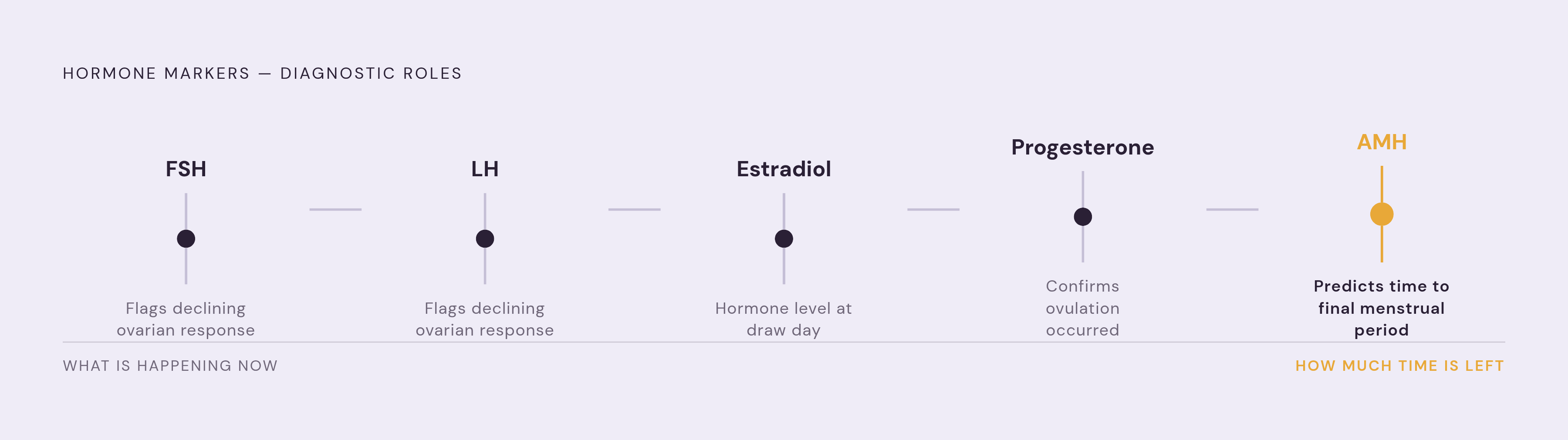
Taken together, these five markers do different jobs: FSH and LH flag that ovarian response is changing, estradiol shows the hormone's actual level on a given day, progesterone confirms ovulation, and AMH projects a timeline forward. Leaving any one of them off a panel means losing a piece of the picture that the others cannot replace. The same logic, it turns out, applies just as forcefully to the thyroid.
Thyroid markers beyond TSH: why TPOAb, Free T3, and Free T4 belong on the list
TSH is the thyroid test almost everyone gets, and it catches the problem most relevant to women moving through perimenopause least often. Autoimmune thyroid disease can be active for years before TSH shows any deviation at all, and by the time it does, the resulting symptoms are hard to separate from the transition already underway.
Thyroid autoimmunity tends to peak during the same decade that perimenopause occupies, and standard TSH screening may not register the shift until thyroid function has already started to change, well after the antibodies driving that change first appeared. A meaningful share of women with a completely normal TSH test positive for thyroid peroxidase antibodies. A woman's TSH can look entirely unremarkable while her thyroid is already under autoimmune attack. That is a diagnostic blind spot, not a minor technicality.
Both an underactive and an overactive thyroid can produce fatigue, mood disturbance, weight changes, sleep disruption, menstrual irregularity, and cognitive difficulty, the exact cluster of symptoms women are told to expect from perimenopause itself. Perimenopause and Hashimoto's thyroiditis overlap so closely on fatigue, brain fog, mood swings, and weight gain that telling them apart requires laboratory data. Free T3 and Free T4 fill in what TSH cannot, since they measure active thyroid hormone circulating in the body rather than the pituitary signal that regulates thyroid output. Including them gives a fuller picture when TSH sits at the edge of normal or when symptoms persist despite a TSH result that looks fine on paper.
Metabolic markers: lipids, glucose, insulin resistance, and estrogen's decline
Estrogen's decline rewrites a woman's metabolic profile even when nothing about her diet or exercise habits has changed, and that is precisely the window these markers exist to catch. Lower estrogen during the menopausal transition has been linked to higher total cholesterol, lower HDL, higher blood pressure, and greater insulin resistance, changes that can appear in women whose lipid profiles looked perfectly healthy before the transition started. A 2025 study measuring physiological markers across a cohort of perimenopausal and postmenopausal women found significant differences in total cholesterol and LDL among women in different age groups spanning the transition, which is the clearest argument for treating a lipid panel as something tracked over years.
A 2026 prospective analysis of postmenopausal women from the UK Biobank, which profiled plasma metabolites by nuclear magnetic resonance and followed participants for over a decade, established that tracking metabolic markers against time since menopause carries real predictive power for cardiovascular events later on. That finding reframes the annual lipid panel as a longitudinal record tracked across years. Non-HDL cholesterol and ApoB are gaining recognition as cardiovascular risk markers that outperform the standard lipid panel, particularly when triglycerides run high or metabolic syndrome is already present, and both belong alongside total cholesterol and LDL.
Glucose regulation shifts in step with estrogen decline, turning insulin resistance into a systemic concern during this life stage rather than something to watch for only in women already flagged as high-risk. HbA1c reflects average glucose over roughly three months, which makes it far less vulnerable to single-day variation than a fasting glucose test taken in isolation. Fasting insulin, read alongside HbA1c, can flag insulin resistance at a point when fasting glucose still reads normal, catching a signal that a standard panel, built around glucose and HbA1c alone, would miss. A meta-analysis of randomized controlled trials found that menopausal hormone therapy significantly reduced insulin resistance in healthy postmenopausal women, a finding that underscores why identifying insulin resistance early, while the transition is still underway, matters for the range of options a woman and her doctor have to choose from.
Ferritin, vitamin D, and hsCRP: the markers that explain symptoms standard testing misses
Fatigue, brain fog, and low mood get attributed to perimenopause by default, when sometimes the actual cause is a nutrient deficiency or an inflammatory marker that was never checked. Ferritin is the clearest example. Low ferritin signals iron deficiency even when hemoglobin reads normal, and a woman can be fatigued, foggy, and unable to sustain exercise without ever meeting the threshold for anemia on a standard CBC. Ferritin also rises during inflammation, so it needs to be read alongside hsCRP to tell true iron depletion apart from an acute-phase response that is masking genuinely low stores.
Vitamin D deficiency is common enough, and symptomatic enough, that its absence from a standard panel is hard to justify. Fatigue, mood disturbance, and musculoskeletal discomfort overlap directly with the symptoms attributed to perimenopause, so checking 25-OH vitamin D rules out a confounding cause. Vitamin D also plays a role in bone metabolism, which matters more as estrogen-related bone loss accelerates through the transition, a connection the next section builds on directly.
hsCRP measures low-level systemic inflammation that standard CRP testing is not sensitive enough to catch. It works as a cardiovascular risk marker in its own right, and it also helps make sense of other results, elevated ferritin being the clearest example of a value that means something different depending on whether inflammation is present. Estrogen carries anti-inflammatory properties, so its decline can shift a woman's inflammatory baseline over the course of the transition, and tracking hsCRP over time makes that shift visible.
Bone turnover markers: why blood-based detection matters before a DXA scan is indicated
Bone loss accelerates during perimenopause itself, well before most women are offered a DXA scan, since DXA scans are typically ordered after menopause rather than during the years when bone density is already declining. A 2025 cross-sectional study in Frontiers in Endocrinology found that bone loss accelerates during perimenopause while DXA remains too insensitive to catch early bone loss, concluding that an easy, sensitive method is urgently needed to identify high-risk women before the damage becomes irreversible.
A blood-based marker is emerging to fill that gap. β-CTX (beta C-terminal telopeptide), paired with estradiol, captures active bone resorption as it happens rather than the cumulative structural loss that a DXA scan reflects only after the fact. Osteoporosis incidence rises after menopause and tracks with age, but the point to intervene is during the transition itself, while bone turnover can still be influenced, not years later once density has already dropped. Muscle mass follows a similar arc, declining at a meaningful rate from the fourth decade onward and accelerating sharply after menopause as estrogen falls, which ties bone health and body composition together as two expressions of the same underlying hormonal shift. Catching that shift through blood markers, rather than waiting for a DXA scan to confirm what has already happened, is what turns bone health from a postmenopausal diagnosis into a perimenopausal opportunity.