Bone Density Baseline Timing for Perimenopausal Women
Baseline bone scans work best before perimenopause's steepest losses.
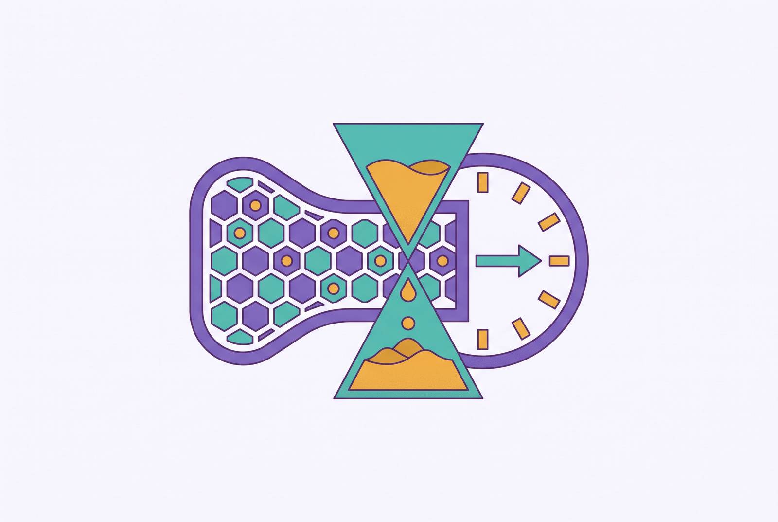
A bone density baseline is most useful before the accelerated loss of late perimenopause, because it lets later measurements show personal change. Longitudinal perimenopause hormone tracking can add context, especially when estradiol trends are interpreted with bone turnover markers and clinician review. Together, these measurements help women and clinicians make decisions sooner with more information, though hormone testing cannot replace a DXA scan.
Why the perimenopausal years are when bone loss is steepest — and hardest to see coming
Bone reaches its peak mass in the second or third decade of life, but its sharpest adult decline is concentrated much later. The Study of Women's Health Across the Nation found that women may lose 5–10% of bone mineral density in cortical and trabecular bone during late perimenopause and the first postmenopausal years. After that window, loss slows to a much lower annual rate. The pattern is therefore not a steady slide through older age. It is an accelerated phase followed by a slower chronic one.
Estrogen helps explain that change of pace. It maintains the balance between bone formation and resorption, the process through which bone is broken down. When estrogen becomes deficient, that balance tips toward resorption, with more breakdown and less formation. This imbalance may already be developing before a woman has reached menopause. Waiting for the transition to feel complete can mean overlooking the years when the skeleton is changing fastest.
Why perimenopause is not a moment but a multi-year hormonal progression, and why that changes what "early" means for bone
That imbalance builds as hormones change over several years. The STRAW+10 framework distinguishes early perimenopause, Stage -2, from late perimenopause, Stage -1. In early perimenopause, progesterone is already declining while estrogen may remain relatively stable or fluctuate high. This pattern, sometimes described as “relative estrogen dominance,” helps explain why symptoms can appear before a woman associates the transition with estrogen deficiency or bone risk. An apparently reassuring estrogen result does not describe the whole progression.
In late perimenopause, estrogen becomes highly erratic, FSH rises, and an absence of periods for 60 or more days is a common marker. An early baseline therefore needs to come before that loss speeds up, which can happen before the final menstrual period. Age cannot reliably locate that window, because the same hormonal shift can arrive in the early 40s or closer to 50. Menstrual patterns and hormonal context matter more to the timing argument than a birthday alone.
What the current screening guidelines actually recommend, and where they leave perimenopausal women
The USPSTF 2025 updated recommendations call for bone density screening in women 65 and older and in postmenopausal women under 65 who have increased fracture risk. For the younger group, the framework for identifying osteoporosis and fracture risk first identifies the presence of 1 or more risk factors, then uses a formal clinical risk assessment tool. It is not a routine baseline programme for the menopausal transition.
The National Osteoporosis Foundation and the International Society of Clinical Densitometry also recommend DXA for postmenopausal women under 65 and women in the menopausal transition who have clinical risk factors for fracture. That transition provision matters, because perimenopausal women are not categorically excluded. Nevertheless, women who still menstruate irregularly and lack those risk flags may receive no prompt for measurement. This leaves a gap between a stage when bone loss can speed up and screening rules that depend on age, postmenopausal status or a qualifying risk factor. It reflects the purpose and architecture of screening.
Why a baseline measurement taken before peak loss has different value than one taken after
An earlier baseline records where a woman's bone density starts before the steepest expected loss, so later measurements can show how it changes. A first scan after that window still shows her current density. What it cannot show is how much density she had before the transition or how quickly she reached the present result. That distinction is the affirmative case for earlier measurement.
The Kuopio OSTPRE population-based cohort study, published in JBMR, followed women with DXA measurements over 25 years. It found that women with higher baseline bone mineral density lost a greater share of it over time. A relatively reassuring later result can therefore coexist with substantial loss from a higher starting point. The study supports attention to personal change, although its long follow-up does not isolate every change to perimenopause. A snapshot and a trajectory answer different clinical questions.
Gourlay et al.'s NEJM study further illustrates how strongly starting density affects decisions about follow-up. In older postmenopausal women, testing intervals ranged from 16-plus years for normal BMD to 1 year for advanced osteopenia. Those findings apply specifically to an older population. Their relevance is that the starting measurement changes the meaning of what comes next.
What DXA actually measures and what a T-score does and does not tell you
DXA is the clinical standard for measuring bone mineral density. It uses a low-radiation X-ray to assess mineral content and produces measurements that support classification as normal, osteopenic or osteoporotic. A T-score shows how density compares with the average peak density in a young adult reference population. T-scores below -2.5 define osteoporosis, while scores between -1.0 and -2.5 define osteopenia.
The practical distinction is between a population benchmark and a personal baseline. A single T-score does not directly measure fracture risk, reveal the speed of bone loss or show the distance from an individual's own peak. Two bone density measurements over time allow a clinician to assess change. That makes the question of how a result compares with the previous scan as important as the category printed on the report. A baseline is useful because it makes that comparison possible.
DXA also has limited sensitivity for early bone loss. Bone breakdown may be increasing even when a scan has yet to show a clear drop in density. This limitation strengthens the case for establishing a reference before acceleration, rather than expecting imaging to identify its earliest biological signs. It also creates a role for measurements that look at bone turnover.
How blood-based markers of bone turnover can flag risk before a DXA scan shows damage
β-CTX, or C-terminal telopeptide, is a blood-based marker of bone resorption, so high levels are a sign that more bone is being broken down. Estradiol provides information about the hormonal setting in which that turnover is occurring. Measuring both asks whether a woman has evidence of increased resorption together with low estrogen. Neither measurement is a substitute for bone density itself.
Feng, Xiao, and Zhang's 2025 study in Frontiers in Endocrinology found significant correlations between estradiol, β-CTX and T-scores in perimenopausal women. Low estradiol combined with elevated β-CTX identified high-risk women with better sensitivity and specificity than either marker alone. Because the study was cross-sectional, it supports risk identification rather than proving that the combination predicts an individual's future loss. Even with that distinction, the finding offers a basis for earlier triage. Blood results may help a clinician decide whose baseline DXA deserves greater urgency.
This lets a woman check her bone health alongside hormone testing she may already be thinking about. Intermission Health's blood panel and clinician review model fits that layered approach through estradiol tracking over time, with β-CTX added to inform the bone resorption picture. Its role is context and triage. For a woman asking how to monitor hormones across multiple months, the useful criterion is a longitudinal record that receives clinical interpretation. The information here does not say how Intermission Health collects samples at home or how often it tests progesterone.
Other factors that affect bone density during perimenopause and should be part of any baseline conversation
Estrogen is central to the timing argument, but it is not the only influence on bone density. Tang et al.'s 2025 study in PeerJ found that visceral fat area was inversely correlated with bone density in perimenopausal women. Women with more visceral fat had lower BMD, while total body fat percentage showed no significant difference. Visceral fat tends to increase during this transition, partly as estrogen declines.
Thyroid function also needs to be considered, though it cannot explain the whole transition. Both overt and subclinical hypothyroidism are more common during perimenopause, and thyroid dysfunction can complicate the assessment of bone turnover. Fatigue, brain fog, mood changes, weight gain and irregular periods can also make it hard to tell thyroid problems from perimenopausal symptoms. Including TSH in a baseline workup helps address that overlap without assuming every symptom is reproductive in origin. The bone conversation should also cover glucocorticoid use, low body weight, smoking, rheumatoid arthritis and diabetes, particularly type 1 diabetes. These factors can compound risk that estradiol alone will not capture.
Why perimenopausal women frequently do not get a bone baseline even when they should
The missed-baseline problem is not simply a shortage of public awareness. Routine screening is more likely to be prompted by a specific risk flag than by a change in hormonal stage. At the same time, only a minority of U.S. OB-GYN residency programs include a dedicated perimenopause and menopause curriculum. That lack of training helps explain why the transition may pass without a discussion of bone health. A consultation organised around current symptoms can leave future skeletal change outside its frame.
Burgin et al.'s 2025 qualitative study in the British Journal of General Practice documented related barriers in perimenopause consultations. Women were uncertain whether their mental health symptoms were connected to perimenopause and sometimes felt inhibited or embarrassed about raising the subject. GPs acknowledged training gaps, while time constraints and stigma further hindered discussion. The study helps explain the consultation context in which a baseline request must compete for attention. When cultural expectations also emphasise hot flashes and fertility rather than bone health, neither the patient nor the clinical pathway may bring the subject forward. Understanding that gap makes self-advocacy more specific.
What a perimenopausal woman can do to get a meaningful bone baseline, and what to ask for
Irregular cycles provide a reason to discuss whether the early perimenopausal window is an appropriate time for a personal bone density reference. The request can be framed as a baseline DXA during the transition, rather than only as screening for established osteoporosis. That distinction makes clear that the clinician is being asked to consider the value of tracking change before the steepest loss.
The same conversation can cover estradiol and FSH for hormonal context, β-CTX for bone resorption risk, and TSH for possible thyroid dysfunction. If a DXA referral is declined because routine guidelines do not require it, relevant personal risk factors may change the assessment. These include low body weight, family history and long-term medication use when they apply to the woman. A bone turnover marker panel can also be requested as a documented first step. The aim is to build a clinically interpreted record.
Follow-up gives that record its value. Two bone density measurements separated by one to two years can reveal change, with the timing discussed in the context of the individual's results and risk. Intermission Health's curated hormone panel, clinician review and longitudinal tracking can add hormonal context between scans, with bone resorption markers contributing another layer where included. Measurement should remain connected to modifiable risk, including calcium and vitamin D intake, weight-bearing exercise, avoiding smoking and avoiding heavy alcohol. The value of a perimenopausal baseline is agency through personal data that support informed decisions with a clinician and put a history behind a first postmenopausal T-score.
Sources
- Osteoporosis Screening Guidelines: Should You Get Tested?
- To scan or not to scan? DXA in postmenopausal women
- A threshold of β-CTX (0.3 ng/mL) with low estradiol identifies high-risk perimenopausal women for bone loss: a cross-sectional study
- Association between visceral fat and bone mineral density in perimenopausal women
- Bone-Density Testing Interval and Transition to Osteoporosis in Older Women